
Polypeptide Structure: Backbone and Folding Levels
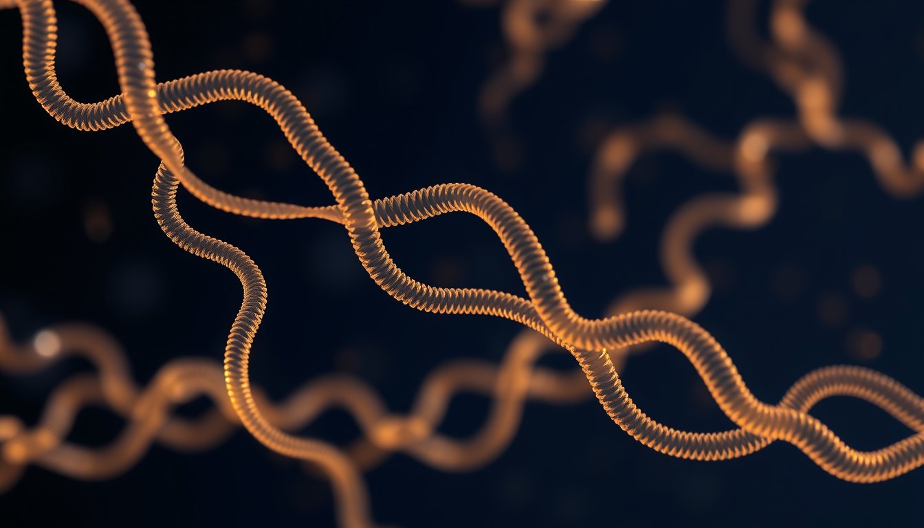
A polypeptide is a chain of amino acids joined by peptide bonds. Its backbone repeats the same three atom unit, nitrogen then alpha carbon then carbonyl carbon, while chemically varied side chains branch off that spine. Biochemists describe the resulting architecture in four levels: primary sequence, secondary structures such as helices and sheets, tertiary three dimensional folding of one chain, and quaternary assembly of several chains. This page summarises what published structural studies describe and gives no health or usage guidance.
A polypeptide is a chain built from amino acid units linked end to end by peptide bonds. The simplest way to picture it is a beaded string: the string itself is chemically identical all the way along, and the beads hanging off it are what differ. Biochemists call the string the backbone (also called the main chain) and the beads the side chains. Almost everything a protein does traces back to how that uniform backbone folds and how the varied side chains are arranged in space once it has folded.
This page covers only the backbone and the four classical levels of structure. Residue numbering conventions and the chemistry of the amino and carboxyl ends of a chain are handled in a separate guide on the polypeptide chain and are not repeated here. This page is for educational purposes only and is not medical advice; consult a licensed physician with any personal health question.
The backbone: a repeating N-C-C unit
Every amino acid residue contributes exactly three atoms to the main chain, always in the same order. Those atoms are the amide nitrogen (N), the alpha carbon (often written C alpha), and the carbonyl carbon (C). Linking residue after residue produces the repeating pattern N-C-C-N-C-C-N-C-C along the entire length of the molecule. This repeat is what people mean by the phrase polypeptide backbone, and the three atom set plus its attached hydrogen and oxygen is sometimes called the peptide group.
Three features of this repeat matter for folding:
- The peptide bond is rigid and planar. The bond joining one residue's carbonyl carbon to the next residue's nitrogen has partial double bond character, so the six atoms around it tend to lie in one plane. Rotation is therefore restricted at that link.
- Rotation happens at the alpha carbon. The two bonds flanking each alpha carbon can rotate, and those two torsion angles, conventionally named phi and psi, define the local shape of the chain. Folding is largely the question of which phi and psi values each residue adopts.
- The backbone carries hydrogen bond donors and acceptors. Each peptide group offers an N-H donor and a C=O acceptor. A review of secondary protein structure described how regular hydrogen bonding between these backbone groups stabilises alpha helices and beta sheets independently of which side chains are present (PMID 29262225).
Side chains: where chemical variety comes from
Attached to each alpha carbon is a side chain, the group that distinguishes one amino acid from another. Side chains range from a single hydrogen atom in glycine to bulky aromatic rings, charged groups, and sulfur containing groups. Because the backbone repeat is constant, side chain identity and order supply essentially all of the chemical information in the chain.
Side chains are also what structural biologists track experimentally alongside the backbone. A 2024 assignment study reported backbone and methyl side chain nuclear magnetic resonance resonance assignments for the Fab fragment of the antibody adalimumab, treating main chain and side chain signals as separate but complementary sets of information (PMID 38926254). Side chains can also be chemically decorated after synthesis: researchers using cell free protein synthesis reported directed glycoengineering of active erythropoietin, a case in which sugar groups attached to the folded chain rather than the backbone atoms themselves were the engineering target (PMID 29867209).
The four levels of structure
| Level | What it describes | Main stabilising interactions |
|---|---|---|
| Primary | The linear order of amino acid residues in one chain | Covalent peptide bonds along the backbone |
| Secondary | Local repeating backbone shapes such as alpha helices and beta sheets | Hydrogen bonds between backbone N-H and C=O groups |
| Tertiary | The full three dimensional fold of a single chain | Side chain packing, hydrophobic burial, salt bridges, disulfide bonds |
| Quaternary | Assembly of two or more folded chains into one functional unit | Interface contacts between separate chains, sometimes disulfide links |
Primary structure
Primary structure is simply the sequence: which residue sits at each position along the backbone, read in a fixed direction. It is the only level defined purely by covalent connectivity. Everything else follows from it in a given environment, which is why sequence changes are the standard lever in protein engineering. A computational design tool described in a 2020 report, IPRO+/-, extended design beyond substitutions by allowing insertions and deletions, meaning the backbone length itself could be altered during redesign (PMID 32857964).
Secondary structure
Secondary structure describes short stretches of backbone that settle into regular, repeating geometries. The two best known are the alpha helix, in which the chain coils so that backbone hydrogen bonds form between residues spaced a few positions apart, and the beta sheet, in which extended strands line up side by side and hydrogen bond across. A review of secondary protein structure set out how these motifs arise from backbone hydrogen bonding patterns and how turns and loops connect them (PMID 29262225).
Beta sheets can also close into cylinders. A methods chapter described the analysis of transmembrane beta barrel proteins by native and semi-native polyacrylamide gel electrophoresis, an approach that distinguishes folded barrel states from unfolded chains (PMID 38478276).
Tertiary structure
Tertiary structure is the complete three dimensional arrangement of one polypeptide chain, including how distant parts of the sequence come into contact. Hydrophobic side chains tend to be buried away from water, polar and charged groups tend to face outward, and covalent cross links can lock parts of the fold together. A 2020 analysis estimated the number of possible disulfide bonded states of native proteins using probabilities of disulfide bond formation, illustrating that even a fixed sequence can in principle pair its cysteines in many different ways (PMID 33291690).
Tertiary architecture also determines catalytic function, because active sites are assembled from residues that are far apart in sequence but adjacent in space. A 2025 preprint described the computational design of cysteine proteases, work that depended on positioning catalytic residues precisely within a designed fold (PMID 41332739); preprint status means the report had not completed peer review at the time of posting.
Quaternary structure
Quaternary structure applies only when two or more folded chains associate into a single functional assembly. The chains may be identical or different, and the interface between them is built from the same kinds of contacts that stabilise tertiary structure. Antibody fragments are a familiar multi chain example, and the 2024 assignment study noted above reported backbone and methyl side chain resonances for the Fab fragment of adalimumab as an intact fragment rather than as isolated chains (PMID 38926254).
Doing the math on a vial? The PeptideU app does reconstitution, units and dilution for you.
Try it freeBackbone mobility and disorder
Not every backbone settles into one fixed shape. Some regions, and some whole proteins, sample many conformations and are described as intrinsically disordered. Modelling such chains accurately has been a persistent technical problem because force fields tuned for folded structures can over stabilise helices. The 2017 CHARMM36m report described a revised force field intended to improve the accuracy of generated backbone conformational ensembles for intrinsically disordered peptides and proteins while preserving performance on folded proteins (PMID 27819658).
Experimental work has measured this mobility directly. A 2018 study of the yeast GRASP protein Grh1 reported high polypeptide backbone mobility together with amyloidogenic behaviour, linking a flexible main chain to a tendency to aggregate (PMID 30356074).
Misfolding and aggregation: what studies report
When backbone hydrogen bonding patterns reorganise across many molecules, chains can stack into ordered aggregates called amyloid fibrils, which are rich in beta sheet. Researchers have developed ways to watch this inside cells: a 2021 study reported Raman spectral imaging of alpha synuclein amyloid fibrils labelled with carbon 13, deuterium and nitrogen 15 in cells, using isotope labelling to separate the fibril signal from the cellular background (PMID 33418468). The Grh1 study described above reported amyloidogenic behaviour for a protein with an unusually mobile backbone (PMID 30356074). These are laboratory and cell level observations about protein behaviour; nothing here describes a treatment, an outcome in people, or any use of a compound.
Tracking research? Log entries with dates, lots and notes: records, never plans.
Get the appRearranging the backbone in the laboratory
Because the backbone is covalent, altering it requires chemistry or enzymology rather than folding alone. Several published approaches target the main chain directly:
- A 2024 report described a chemical counterpart to the resolution step of nature's intein mediated protein splicing, mimicking the reaction by which inteins excise themselves and rejoin the flanking chain (PMID 38096499).
- A 2024 study reported sortase catalysed protein domain inversion, an enzymatic strategy that changed the order of domains along a polypeptide chain (PMID 38366985).
- The IPRO+/- design tool was reported to permit insertions and deletions during computational redesign, changing backbone length rather than only side chain identity (PMID 32857964).
Common points of confusion
- Backbone versus side chain. The backbone is the invariant N-C-C repeat; side chains are the variable groups attached at each alpha carbon. Nuclear magnetic resonance studies routinely assign the two separately (PMID 38926254).
- Secondary structure is not a fold. Helices and sheets are local backbone geometries held by main chain hydrogen bonds (PMID 29262225); the complete fold is tertiary structure.
- Quaternary structure requires more than one chain. A single long chain folded into several domains still has tertiary, not quaternary, structure.
- Disorder is not an error. Flexible backbone ensembles are a recognised modelling and measurement challenge rather than an artefact, as the CHARMM36m report described (PMID 27819658).
Want the full course? Every compound, evidence-graded and cited, inside PeptideU.
Start learning freeLimits of what this summary covers
The studies summarised here are structural, computational and methodological. They describe how chains are built, measured, modelled and rearranged in laboratory settings. None of them is cited on this page as evidence about safety, benefit or use of any substance in people, and no dose or treatment information appears anywhere on this page. Readers with a personal health question should raise it with a licensed physician.
References
- Biochemistry, Secondary Protein Structure (StatPearls, 2022)
- CHARMM36m: an improved force field for folded and intrinsically disordered proteins (Nature Methods, 2017)
- Backbone and methyl side-chain resonance assignments of the Fab fragment of adalimumab (Biomolecular NMR Assignments, 2024)
- Multiple Disulfide-Bonded States of Native Proteins: Estimate of Number Using Probabilities of Disulfide Bond Formation (Molecules, 2020)
- Analysis of Transmembrane beta-Barrel Proteins by Native and Semi-native Polyacrylamide Gel Electrophoresis (Methods in Molecular Biology, 2024)
- The yeast GRASP Grh1 displays a high polypeptide backbone mobility along with an amyloidogenic behavior (Scientific Reports, 2018)
- Raman spectral imaging of 13C 2H 15N-labeled alpha-synuclein amyloid fibrils in cells (Biophysical Chemistry, 2021)
- A Chemical Counterpart to the Resolution Step of Nature's Intein-Mediated Protein Splicing (ACS Chemical Biology, 2024)
- Sortase-Catalyzed Protein Domain Inversion (Angewandte Chemie International Edition, 2024)
- IPRO+/-: Computational Protein Design Tool Allowing for Insertions and Deletions (Structure, 2020)
- Cell-free protein synthesis as a novel tool for directed glycoengineering of active erythropoietin (Scientific Reports, 2018)
- Computational design of cysteine proteases (bioRxiv preprint, 2025)
Frequently asked questions
What is the polypeptide backbone?▾
The backbone is the continuous chain of atoms shared by every amino acid residue: an amide nitrogen, an alpha carbon, and a carbonyl carbon, repeated as N-C-C along the molecule. Its N-H and C=O groups form the hydrogen bonds that stabilise alpha helices and beta sheets, as described in a review of secondary protein structure (PMID 29262225).
What is a peptide group?▾
The peptide group is the planar unit formed around the bond linking one residue's carbonyl carbon to the next residue's nitrogen, including the attached oxygen and hydrogen. Its rigidity restricts rotation, leaving the two torsion angles at each alpha carbon to define local shape. A review of secondary structure described how these backbone groups hydrogen bond into regular motifs (PMID 29262225).
How do the four levels of structure differ?▾
Primary structure is the residue sequence, secondary structure is local backbone geometry such as helices and sheets (PMID 29262225), tertiary structure is the full three dimensional fold of one chain, and quaternary structure is the assembly of several chains. A 2024 study reported backbone and methyl side chain assignments for the multi chain Fab fragment of adalimumab (PMID 38926254).
Do all polypeptides fold into one fixed shape?▾
No. Some chains sample many conformations and are called intrinsically disordered. The 2017 CHARMM36m report described a revised force field aimed at generating more accurate backbone ensembles for intrinsically disordered proteins while preserving folded protein accuracy (PMID 27819658). A 2018 study reported high backbone mobility in the yeast GRASP protein Grh1 (PMID 30356074).
What role do side chains play compared with the backbone?▾
The backbone repeat is chemically identical in every residue, so side chains supply the variation that drives packing, charge and catalysis. Side chains can also be modified after synthesis: researchers reported directed glycoengineering of active erythropoietin using cell free protein synthesis (PMID 29867209), and a 2025 preprint described computational design of cysteine proteases (PMID 41332739).
Can the backbone itself be rearranged in the laboratory?▾
Published work describes several approaches. A 2024 study reported sortase catalysed inversion of domain order within a polypeptide chain (PMID 38366985), and another 2024 report described a chemical counterpart to the resolution step of intein mediated protein splicing (PMID 38096499). A design tool was also reported to allow insertions and deletions that change backbone length (PMID 32857964).
How do misfolded backbones form amyloid fibrils?▾
When many chains adopt extended beta sheet geometry and stack together, ordered fibrils can form. A 2021 study reported Raman spectral imaging of isotope labelled alpha synuclein amyloid fibrils inside cells (PMID 33418468), and a 2018 study reported amyloidogenic behaviour alongside high backbone mobility in Grh1 (PMID 30356074). These are laboratory observations, not health guidance.
Track it. Calculate it. Actually understand it.
References
This page summarises published research for education. It is not medical advice, and nothing here is a recommendation to use, purchase, or dose any substance. Study parameters described are what researchers reported, not instructions. Consult a qualified clinician before any health decision.